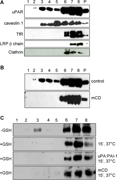
Figure 5. Role of Lipid Rafts in uPA:PAI-1 dependent and ligand-independent uPAR endocytosis.
Panel A: Flotation assay of TX-100 extracts of HEK293-uPAR analyzed by immunoblotting. uPAR is located in both detergent-resistant (fraction 2 and 3) but mostly in detergent-soluble fractions of the gradient (lanes 6–8). Caveolin-1 displays a similar distribution, while TfR, LRP-1, β1 integrin and EEA1 are exclusively found in the detergent-soluble fractions. P: pellet. Panel B: uPAR flotation in control and beta-methyl-cyclodextrin (10 mM, 30 minutes at 37°C) treated cells. Under these conditions, uPAR is found only in detergent-soluble fractions (Lanes 6–8). Notice that the flotation in untreated cells is the same as in Panel A. Panel C: flotation analysis of uPAR internalization with cell surface biotinylated HEK293-uPAR cells. UPAR is located in lipid rafts but it is internalized outside of these microdomains. Upper row: cells treated with biotin but not subjected to reduction with GSH, showing the distribution of uPAR present at the cell surface. Second row: uPAR internalized in the absence of ligands (15 min 37°C). Third row: uPA:PAI-1-dependent uPAR internalization. Fourth row: uPAR constitutively internalized in beta-methyl-cyclodextrin treated cells. All images are representative of 3–4 independent experiments.

