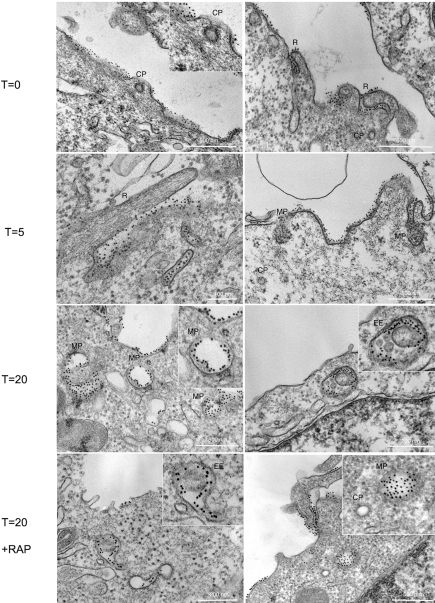
Figure 6. Ultrastructural visualization of uPAR endocytic vesicles.
HEK293-uPAR cells were incubated with anti-uPAR monoclonal antibody R3 (that recognizes the D1 extracellular domain) at 4°C for 20 minutes, washed and then probed with protein A gold 10 nm in the absence and in the presence of 200 nM RAP. After extensive washing, cells were warmed in culture medium for 5 to 20 minutes at 37°C to reveal the identity of uPAR primary endocytic vesicles. Samples were then fixed in 2.5% Glutaraldehyde in 0.1 M Sodium Cacodylate buffer and processed for standard EM plastic embedding. At t = 0, uPAR labelling was observed along the plasma membrane, especially in membrane ruffles (R) but excluded from clathrin coated pits (CP). Upon warming at 37°C, uPAR labelling was observed in ruffling regions of the membrane (R), and in clearly large uncoated vesicular profiles of similar morphology to macropinosomes (MP) at t = 5 and t = 20 minutes as well as in early endocytic elements at 20 minutes. uPAR is visible in endocytic vescicles upon 20 minutes internalization in the presence of 200 nM RAP. Note the full macropinosome (MP) and the empty clathrin-coated pit (CP).

