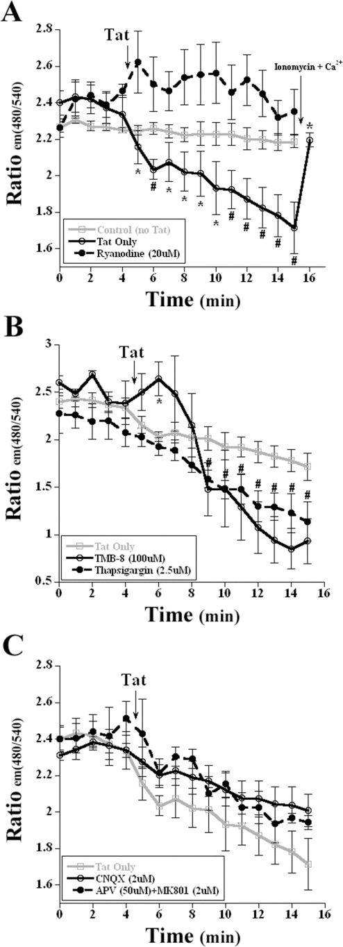
Figure 1. Endoplasmic reticulum calcium decrease via RyR in response to HIV-1 Tat.
A, Application of 8 nM Tat to transfected neurons induced the loss of the CFP:EYFP fluorescent signal, indicating a loss in ER calcium (labeled ‘Tat Only’). Pretreatment with ryanodine for 30 min prior to exposure inhibited this loss in ER calcium. To ensure that the loss in fluorescence was not due to overexposure, a photo-bleaching control, Control (no Tat), was performed to demonstrate the stability of the fluorescent signal. Application of the positive controls, ionomycin (2 µM) or calcium (10 mM) increased the ER CFP:EYFP fluorescence (n = 5; *, p<0.05; #, p<0.01). B, Pretreatment with the IP3 inhibitor TMB-8 and the sarco-endoplasmic reticulum Ca2+-ATPase inhibitor thapsigargin failed to attenuate the loss of ER calcium when the transfected cortical neurons were exposed to 8 nM Tat. (n = 4; *, p<0.05 for TMB-8; #, p<0.05 for thapsigargin). C, Transfected neurons that were pretreated with either APV/MK-801 or CNQX exhibit the same relative loss of the ER CFP:EYFP fluorescence, demonstrating the loss of calcium from the ER and not influx from activation of ionotropic glutamate receptors (n = 4). In Figure 1A, the mean values of CFP:EYFP fluorescence for ‘Tat Only’ treatment group were plotted, and treated time points were compared with control time points (before addition of Tat) for statistical significance. For all other groups (Figure 1A–C), the values of CFP:EYFP fluorescence were plotted and all time points were compared with the treatment group ‘Tat Only’ time points for statistical significance.

