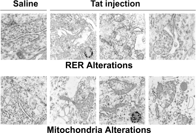
Figure 6. HIV-1 Tat induces endoplasmic reticulum and mitochondrial changes in frontal cortex of mice.
Wild type C57Bl/6J received stereotactic injections of vehicle control or HIV-1 Tat (50 µmols) into frontal cortex, followed by sacrifice 4 weeks later. Frontal cortex was processed for electron microscopy. The upper and lower panels on the left depict normal morphology of RER and mitochondria, respectively. The upper panels on the right demonstrate mild ER dilatation with occasional vacuolization. The lower panels on the right demonstrate abnormally enlarged mitochondria with increased cristae.

