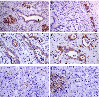
Figure 2.
Immunohistochemical staining of PDAC samples. Moderate KLK6 immunoexpression in pancreatic ducts (arrow) and strong expression in Langerhans' islets (arrowhead) no staining in acini ( × 100) (A). Strong KLK10 immunoexpression in the crypts of the intestinal epithelium of the ampulla of Vater ( × 100) (B). Strong KLK6 immunoexpression in pancreatic adenocarcinomas ( × 200) (C and D). Moderate KLK10 immunoexpression in pancreatic adenocarcinomas ( × 200) (E). Strong KLK10 immunoexpression in Langerhans' islets (arrow), absence of expression in pancreatic adenocarcinoma (arrowhead) ( × 200) (F).

