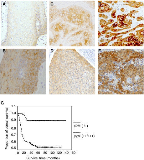
Figure 2.
Immunohistochemical staining for β2M and overall survival in patients with OCSCC. (A–F) The β2M intensity on tissues was evaluated by immunohistochemical staining. (A) The β2M protein was localised at plasma membrane in normal mucosa tissue. Tumour tissues with plasma membrane (C, E) or cytoplasmic (D, F) β2M staining were classified according to a two-grade scale: absent or weak staining (−/+ C, D), and strong staining (++/+++, E, F) as compared with weak β2M staining in adjacent non-tumour oral tissue (B). (G) The survival period for those patients (n=184) with strong (++/+++) tumour expression (dashed line) was significantly shorter than that for those (n=72) with absent or weak (–/+) expression (solid line). The difference in survival was statistically significant (P<0.001) according to the log-rank test. Overall survival was calculated from the time of surgery to the date of death, the event of interest, or the date of last follow-up. All statistical analyses were performed using SPSS software.

