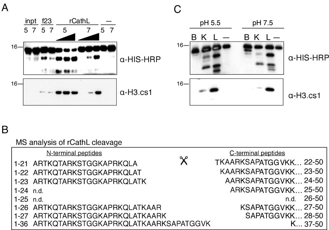
Figure 5. rCathepsin L cleaves histone H3 in vitro.
(A) Recombinant Cathepsin L cleaves rH3 in vitro at both pH 5.5 and pH 7.4 and generates a fragment that is recognized by both α-HIS-HRP (top panel) and α-H3.cs1 (middle panel) antibodies.
(B) Recombinant mouse Cathepsin L was incubated with recombinant H3-HIS at both pH 5.5 and pH 7.4; after 2 hours, the reaction products were subjected to analysis by mass spectrometry. Both N-terminal (left) and C-terminal (right) fragments of the rH3 cleavage were detected; note the similarity to the pattern of in vivo cleavage shown in Figure 2B.
(C) Recombinant Cathepsins B, K, and L were pre-activated and incubated with recombinant H3-HIS at both pH 5.5 and pH 7.4 for 15 minutes; the reactions were separated by SDS-PAGE and analyzed by immunoblotting.

