Fig. 4.
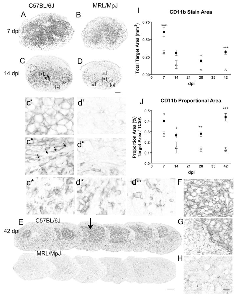
CD11b (Mac-1) immunoreactivity was diminished in MRL/MpJ mice at all time points examined. Corresponding sections from the epicenter of C57BL/6J (A, C, E–G) and MRL/MpJ (B, D, E, H) specimens were stained simultaneously. (A) Macrophages are found throughout the lesion center in both strains at 7 dpi. (B) The area and intensity of Mac-1 staining were lower in MRL/MpJ mice; (C) 14 dpi in C57BL/6J mice. Boxes indicate regions enlarged below. The lesion contains a large area of Mac-1 staining, with rounded MCs throughout the center (c′), and a thin rim devoid of Mac-1 staining bordering the clusters (c″ arrows). Below this rim, activated Mac-1 positive cells exhibit heterogeneous morphologies suggesting a microglial origin. (D) Little to no staining was found in the lesion center in the MRL/MpJ mice at 14 dpi (d″), while elongated Mac-1+ cells line the borders (d″). Both strains have similar microglial distribution and activation in the rim of white matter (c*, d*, d**); (E) 42 dpi. Mac-1-stained sections spaced 300 μm apart through the lesion site in representative animals at 42 dpi illustrating the marked reduction in macrophage staining in MRL/MpJ mice at this chronic time point. The lesion epicenter section is marked by the arrow. (F–H) Enlargements from the sections stained at 42 dpi. (F) Center of a C57BL/6J lesion. (G) Border of the lesion center in C57BL/6J specimen. (H) Center of an MRL/MpJ lesion. (E, I, J) Time course of total (I) and proportional (J) area measures of Mac-1 (CD11b) labeled-macrophage/microglial profiles at the epicenter section in C57BL/6J and MRL/MpJ specimens Two-way ANOVA effect of dpi and strain for both measures (P<0.0001), differences between strains at each time point marked with asterisks (* P<0.05, ** P<0.01, *** P<0.0001). Scale bar=200 μm A–E; d**: Scale bar=10 μm c–d**. Scale bar=50 μm F–H.

