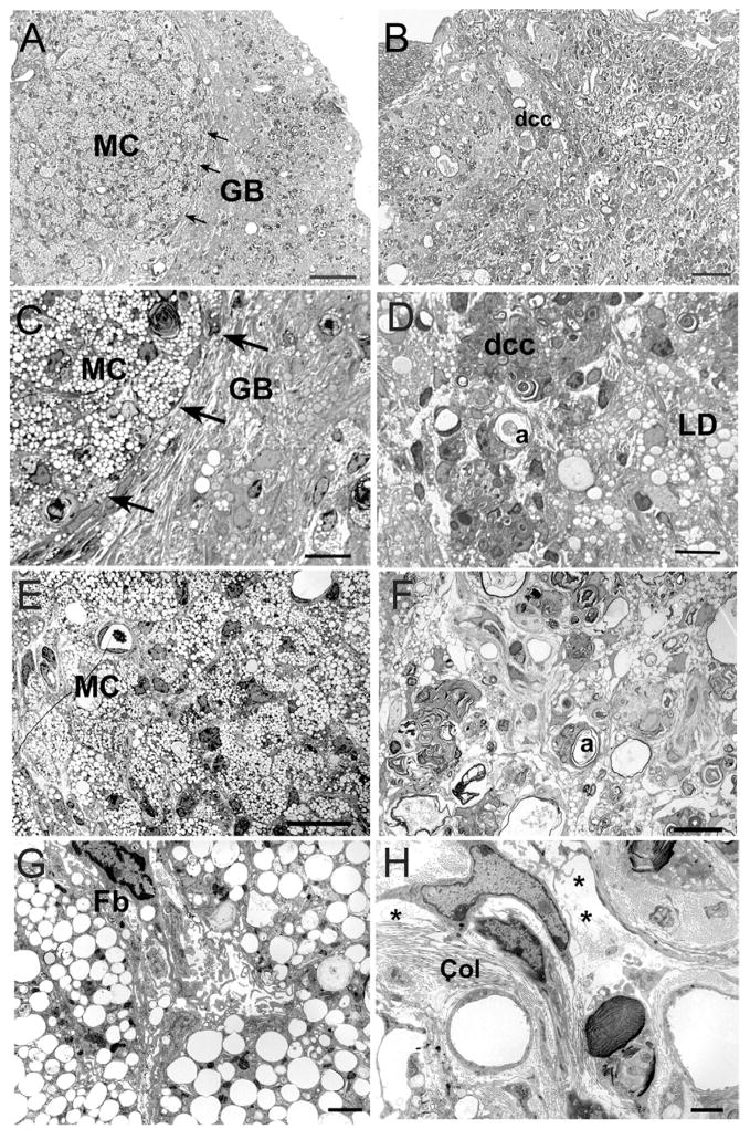
Fig. 6.
Light and electron microscopy through the epicenter reveals a compact composition in C57BL/6J (A, C, E, G) and diffuse cellular organization in MRL/MpJ (B, D, F, H) specimens at 42 dpi. (A–D) Light micrographs from the lateral border of the lesion zones. (A, C) In C57BL/6J mice, packed MC are clearly isolated from a dense glial bundle that surrounds the lesion (GB, arrows). (B, D) The glial border in MRL/MpJ specimens is complex and poorly delineated. MRL/MpJ specimens lack large clusters of macrophages, but contain scattered lipid droplets (LD), degenerating axons (a) and loosely packed tissue elements. Note the presence of clusters of cells with dcc. (E–H) Light (E, F) and electron (G, H) micrographs from the center of the lesion sites. (E, G) C57BL/6J lesions are filled with tightly packed MC and Fb-like cells. (F, H) In MRL/MpJ specimens, degenerating axons (a) are interspersed with lipid droplets (LD) and lightly stained cell clusters, often found surrounding blood vessels. (H) Higher magnification illustrates the presence of abundant extracellular space (*) and numerous collagen fibers (Cl) throughout the MRL/MpJ lesions. Scale bar=50 μm (A, B); 20 μm (C, D); 10 μm (E, F); 2 μm (G, H).

