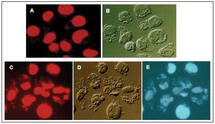
Figure 2.
Untreated (A and B) and TK6 cells treated with 100 µg/ml CPFX for 24 h (C-E) were stained with 7-AAD and examined by microscopy (Nikon Microphot FXA, 60×). The red fluorescence emission of DNA-bound 7-AAD (A and C) was induced by exposure to green wavelengths and the blue emission of CPFX (E) was induced by exposure to UV light. (B and D) show cell morphology under differential interference (Nomarski) contrast. Note extensive nuclear fragmentation and formation of apoptotic bodies (C-E), changes typical of apoptosis, in cells treated with CPFX. Interestingly, the blue fluorescence of CPFX is primarily associated with nuclei and fragments of chromatin

