
Figure 2.
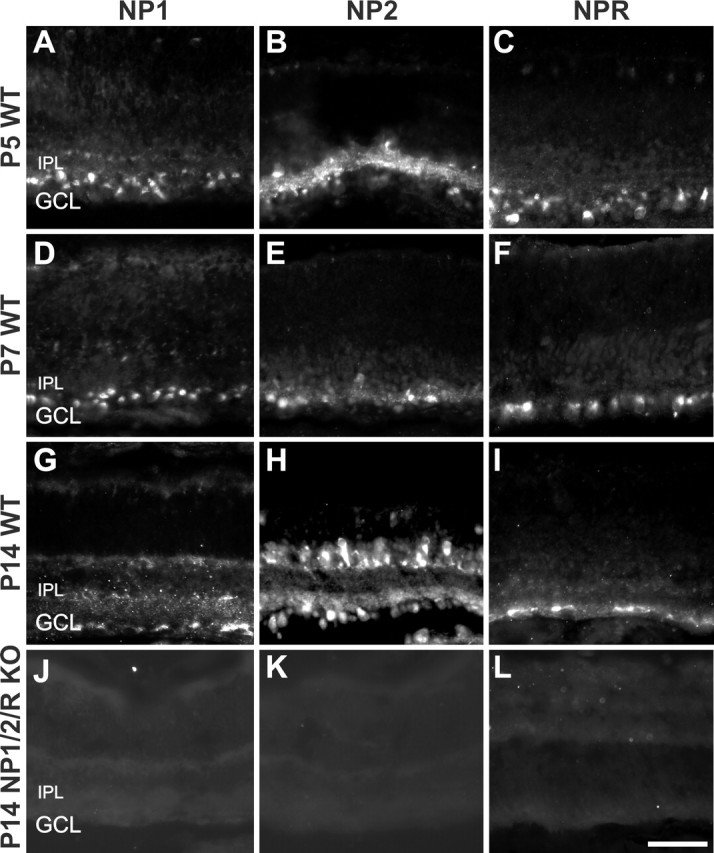
Immunofluorescence images of postnatal mouse retina stained with antibodies to NP1 (first column), NP2 (second column), or NPR (third column) at P5, P7, or P14. A–I, WT retinas. J–L, NP1/2/R KO retinas. In WT retinas (A–I), NP1-, NP2-, and NPR-immunopositive cells are seen in the ganglion cell layer at every age examined. No specific signal is seen in the retinas of NP1/2/R KO mice (J–L). IPL, Inner plexiform layer; GCL, ganglion cell layer. Scale bar, 50 μm.