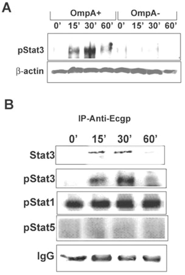
Fig. 1. Association of phospho-Stat3 with Ec-gp96 during the invasion of OmpA+ E. coli in HBMEC.

A. Confluent HBMEC monolayers were infected with either OmpA+ E. coli or OmpA− E. coli for varying periods, washed, and total lysates were prepared. Approximately 20 μg of total lysates was analysed by Western blotting using a phospho-Stat3 antibody. Protein loading in the samples was examined by assessing the amount of β-actin in the lysates.
B. Approximately 300 μg of the lysates was subjected to immunoprecipitation with anti-Ec-gp96 antibody and the resulting immune complexes were analysed for association of phosphorylated Stat1, Stat3 or Stat5. Equality of protein loading in the gel was assessed by the amount of IgG present in each lane.
