
Figure 3. Imatinib mesylate hampers Treg suppressive activity and FoxP3 expression.
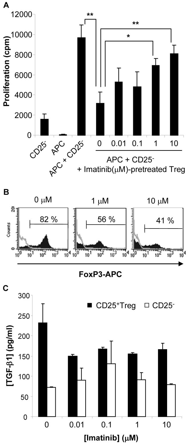
CD4+CD25+ Treg from lymphoid tissues of BALB/c mice were exposed to increasing but non-toxic concentrations of imatinib mesylate for 48 hrs, recovered and washed 3 times in complete medium to eliminate residual imatinib mesylate. (A) The effect of treated or untreated Treg on the proliferation of responder CD4+CD25− T lymphocytes (CD25−) stimulated with mitomycin C-treated allogeneic APC was determined by [3H]-thymidine incorporation assays. *, p<0.02; **, p<0.005. (B) Expression of FoxP3 in CD4+CD25+ Treg treated for 48 hrs with the indicated concentrations of imatinib. Representative results of three independent experiments. (C) TGF-β1 concentration was assessed by ELISA in imatinib mesylate-treated (48 hrs) CD4+CD25+ Treg (CD25+Treg) or CD4+CD25− T cell (CD25−) cultures. No significant difference was found between groups. Representative results of two independent experiments.