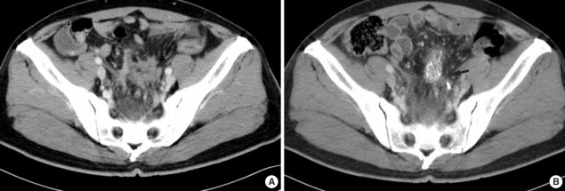
Fig. 2.
Primary sigmoid colon cancer. (A) Before chemotherapy, an exophytic tumor mass (arrow) invading the distal left ureter. (B) After chemotherapy, a slight decrease in the size of the tumor mass (arrow) but no definite interval change. A Hanaro Colorectal Stent® and a double J ureteral stent were visible.

