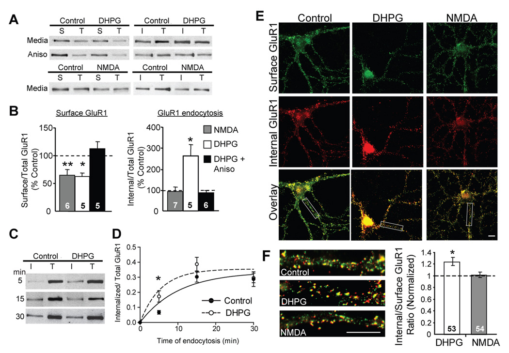
Figure 1. Brief mGluR activation induces a persistent and protein synthesis dependent increase in GluR1 endocytosis rate.
A, Representative blots of surface (S), internalized (I) (100 µg pulldown) and total (T) GluR1 (10 µg protein) from high-density dissociated hippocampal neuron cultures one hour after DHPG or NMDA treatment in media or in the presence of anisomycin (Aniso). B, Brief DHPG treatment (100 µM; 5 min) results in persistent (60 min) decrease in surface GluR1 and increases in endocytosis rate for GluR1 measured with receptor biotinylation relative to control, or media treated, sister cultures. Preincubation of cultures in anisomycin (20 µM) blocks surface GluR1 decreases and GluR1 endocytosis rate increases. Although application of NMDA (20 µM; 3 min) results in a persistent (60 min) decrease in surface GluR1, it does not persistently alter GluR1 endocytosis rate. N = # cultures per condition is on each bar. Paired t-tests were performed on raw ratio values in treated vs. their respective control, media treated cultures (Supp. Table 1). C, Representative western blots of internalized (I) and total (T) GluR1 in high-density hippocampal cultures. One hour after treatment with media or DHPG, surface receptors were labeled with biotin and allowed to internalize at 37°C for 5,15 or 30 minutes, after which the remaining surface biotin was stripped off and internalized, biotinylated GluR1 was measured. D, Quantification of internalized/total GluR1 levels from data in C reveal that GluR1 endocytosis is not saturated at early time points (5 min) in either control or DHPG-treated cultures and therefore reflects endocytosis rate. N = 5 cultures per condition. A best fit single exponential association function (using the Marquardt and Levenberg method) was used to obtain a τ for endocytosis. E, Representative double-label images of surface (green) and internalized (red) staining of GluR1 in low-density hippocampal neurons. Live cultures were labeled with N-terminal GluR1 antibody one hour after treatment with media (Control), DHPG or NMDA. F, Left: Representative proximal dendrites from images of merged surface and intracellular GluR1 immunostaining. Right: Ratiometric analysis of internal to surface GluR1 puncta number reveal that one hour after treatment, DHPG, but not NMDA, persistently increases the internalization rate constant for GluR1. N = # cells per condition is on each bar. Scale bars = 10 µm. Data pooled from 4 cultures each. For all figures * p < 0.05; ** p < 0.01; ***p< 0.001*.

