
Fig. 4.
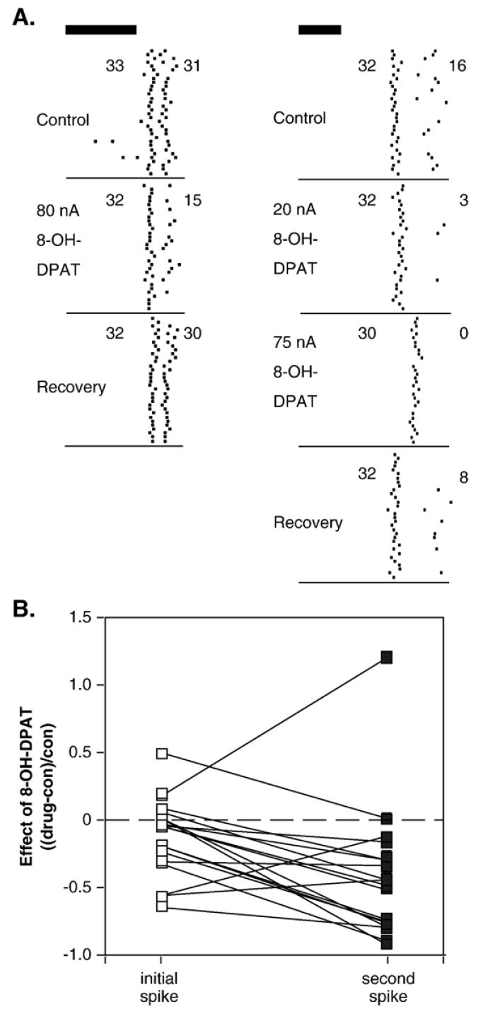
8-OH-DPAT alters the temporal structure of spike trains in single neurons. (A) Peri-stimulus time histograms of two different neurons that both show greater depression of second as opposed to initial spikes in the presence of 8-OH-DPAT. The stimulus presented to the neuron on the left was a 10-kHz FM sweep centered at 24 kHz, at 50-dB SPL, and the stimulus on the right was a 10-kHz FM sweep centered at 24 kHz, at 30-dB SPL. Stimuli for both neurons, represented by dark bars, were 10-ms long. Numbers represent responses for the initial versus second spikes. (B) Effects of 8-OH-DPAT on initial versus second spikes for 18 neurons with consistent intervals between the first and second spike. Values are changes in spike count in the presence of 8-OH-DPAT normalized to the control values for the initial (open symbols) or second (closed symbols) spike. Lines connect values from the same neuron.