
FIG. 4.
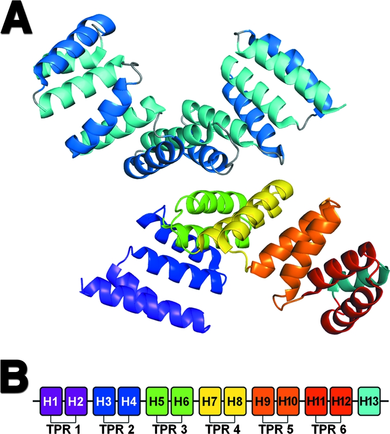
Crystallographic structure of PilF. (A) Ribbon diagram showing the arrangement of the two molecules of PilF present in the crystallographic asymmetric unit. The top molecule has been colored to show the bilayered arrangement of helices. Helices in the inner concave and outer convex surfaces are colored in light and dark blue, respectively. The second (bottom) molecule has been colored to show the six TPR motifs found in PilF. The TPR motifs are colored from purple at the N terminus to red at the C terminus. The final solvation helix is shown in light blue. (B) Schematic representation of the 13 helices found in PilF. The six TPR motifs and C-terminal solvation helix are colored as described for the second (bottom) molecule in panel A.