
FIG. 5.
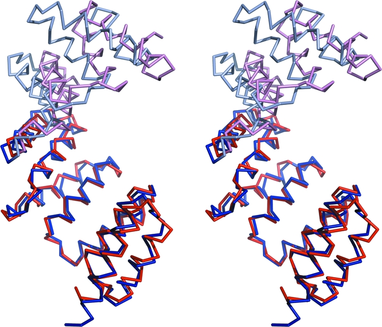
Structural differences between PilF and PilW. Stereo view of the Cα trace of PilW superimposed on PilF. The proteins were superimposed using TPR motifs 1 to 4, comprising residues 34 to 170 in PilF. TPR motifs 1 to 4 of PilF and PilW are drawn in dark blue and dark red, respectively. The remaining regions of PilF and PilW are drawn in light blue and light red, respectively.