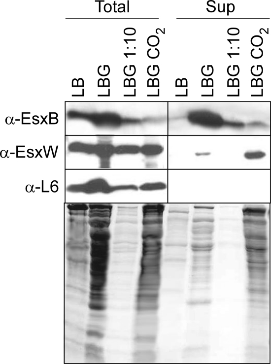
FIG. 2.
Detection and localization of EsxB and EsxW by Western blotting. Cultures of B. anthracis Sterne were grown to A600 nm of 3 in either Luria broth alone (LB) or complemented with glucose 0.5% (LBG) or glucose 0.5% and sodium bicarbonate 0.85% (LBG NaCO2). A volume of 1 ml was precipitated with TCA to examine total proteins in the culture (Total) fraction, while 1 ml of supernatant was recovered after centrifugation of a 3-ml volume culture. Proteins in the supernatant (Sup) fraction were submitted to TCA precipitation. TCA pellets were washed with acetone and solubilized in SDS-PAGE loading buffer before separation on a 15% SDS-PAGE gel. The gels were either stained with Coomassie blue or used for Western blot analysis. The presence of EsxB and EsxW was revealed by immunoblotting using polyclonal antibodies. The ribosomal protein L6 was used as a nonsecreted cytosolic marker. The same sample volumes were loaded in the gels. For quantitative comparison, samples grown in LBG were loaded undiluted and diluted 1:10. α, anti.

