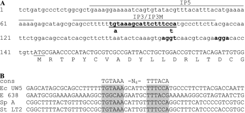
FIG. 1.
(A) Nucleotide sequence of the region upstream of ipdC in E. cloacae UW5 (GenBank accession no. AF285632). The putative TyrR box is indicated in boldface and underlined, the translational start site is underlined, the 5′ partial coding sequence is capitalized, and putative ribosome-binding sites are highlighted in boldface and underlined. The location of IP5, IP3, and IP3M used in EMSAs are overlined. Nucleotide substitutions and locations of mutations present in IP3M are shown below the wild-type IP3 sequence. (B) Alignment of ipdC promoter sequences from E. cloacae UW5 (Ec UW5), Enterobacter sp. strain 638 (E 638), S. enterica subsp. enterica serovar Paratyphi A (Sp A), and serovar Typhimurium LT2 (Sp LT2) (GenBank accession nos. AF285632, CP000653, CP000026, and AE008808, respectively). The consensus sequence for the TyrR binding site in E. coli (cons) is shown. Shaded in gray are the conserved putative TyrR boxes in the ipdC promoter sequences.

