
Figure 6.
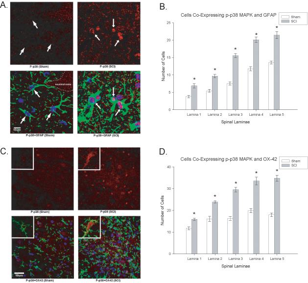
SCI demonstrates higher magnification of increased cellular localization of activated p38 (p-p38, red) in both astrocytes (Figure 6A, GFAP positive, green) and microglia (Figure 6B, OX-42 positive, green). Note that the p-p38 is increased in the nuclear compartment of both cell types. Quantification of the significant increases in p-p38 MAPK in astrocytes (GFAP, 6B) and microglia (OX-42, 6D) in the dorsal horn laminae of SCI rats relative to sham rats (*p<0.001). Quantification of the significant increases in p-p38 MAPK colocalization with GFAP (6B) and OX-42 (6D) positive cells (astrocytes and microglia, respectively) in the dorsal horn laminae of SCI rats relative to sham rats (*p < 0.001). All data are represented as mean ± SEM.