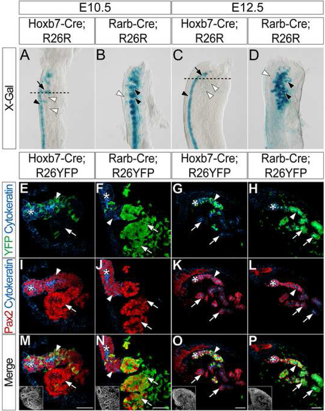
Figure 1. Cranial mesonephric tubules are primarily derived from anterior intermediate mesoderm mesenchyme.
(A–D) X-gal staining in the mesonephros of E10.5 (A) and E12.5 (C) Hoxb7-Cre;R26R or E10.5 (B) and E12.5 (D) Rarb-Cre;R26R embryos. Black arrowheads (A, C) indicate X-gal staining in the ND. Black arrows (A, C) indicate X-gal staining in ND outgrowths. Black arrowheads (B, D) indicate X-gal staining in the mesonephric tubules. White arrowheads (B, D) indicate lack of X-gal staining in the ND. Dashed lines (A, C) indicate approximate planes of section (E–T). (E–T) Immunofluorescent confocal microscopy of transverse sections of anterior mesonephros in E10.5 (E, I, M) and E12.5 (G, K, O) Hoxb7-Cre;R26YFP or E10.5 (F, J, N) and E12.5 (H, L, P) Rarb-Cre;R26YFP embryos stained for YFP, Cytokeratin and Pax2. Insets (M, N, O, P) indicate nuclei. Asterisks indicate the ND. Arrowheads indicate ND outgrowths (E, F, I, J, M, N) or mesonephric tubule connecting segments (G, H, K, L, O, P). Arrows (E–P) indicate developing mesonephric tubules. Scale bars in M, N, O and P = 50µm.

