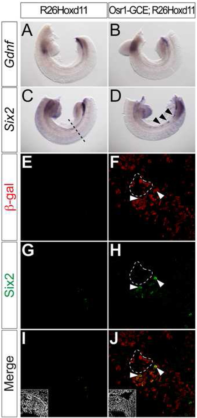
Figure 4. Hoxd11 activates cell-autonomous activation of Six2, but not Gdnf, in mesonephric mesenchyme.
(A–D) Whole mount in situ hybridization for Gdnf (A, B) and Six2 (C, D) in E10.5 control (A, C) or Osr1-GCE;R26Hoxd11 (B, D) embryos. Arrowheads (D) indicate sites of ectopic Six2 expression. Dashed line (C) indicates approximate plane of section (E–L). (E–L) Immunofluorescent confocal microscopy of transverse sections of the mesonephros in E10.5 control (E, G, I) or Osr1-GCE;R26HoxD11 (F, H, J) embryos stained for β-gal and Six2. Arrowheads (F, H, J) indicate examples of β-gal and Six2 co-localization. White dashed line (F, H, J) indicates ND epithelia positive for β-gal, but not Six2. Insets (I, J) indicate nuclei.

