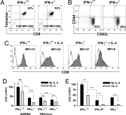
Fig. 1.
CD8 expression by activated CD8+ T cells is reduced in the presence of IL-4 and/or the absence of IFN-γ. CD8+ cells from OT-I (IFN-γ+/+) and OT-I x IFN-γ−/− (IFN-γ−/−) mice were stained with (A) anti-CD8α Ab and SIINFEKL tetramer or (B) Ab to CD44 and CD62L. The CD8 MFI and the percentage of double-positive cells (A), and the percentage of cells with a naïve (CD44low CD62Lhigh) phenotype (B), are shown within the frames. (C) The two CD8+ populations were cultured with anti-receptor Ab and IL-2 with or without IL-4 for 6 days then stained with anti-CD8α Ab (filled histograms) or isotype control Ab (open histograms). (D) The same CD8+ populations were cultured with SIINFEKL-coated APC or PMA and ionomycin, and IL-2 with or without IL-4. CD8 expression is shown as relative fluorescence intensity, obtained by normalizing the MFI to the highest sample in the experiment (100%). (E) CD8 expression by CD8+ cells from C57BL/6 WT (IFN-γ+/+), IFN-γR−/− or IFN-γ−/− mice was measured after culture with anti-receptor Ab, IL-2 and with or without IL-4 for 10 days. Groups were compared by unpaired t test (see Materials and Methods).

