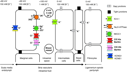
Figure 6.
Schematic model of cochlear potassium secretion across the stria vascularis and of the formation of endocochlear potential (EP). The stria is an unusual multilayered epithelium in which tight junctions between marginal cells, which face directly the endolymph, as well as tight junctions between basal cells isolate an intrastrial space that has a low K+ concentration. The EP is largely generated by a K+-diffusion potential through the Kir4.1 K+ channel across the apical membrane of intermediate cells. The loss of barttin abolishes currents through both ClC-Ka and ClC-Kb, indirectly impairing basolateral K+ uptake by the NaK2Cl cotransporter and the Na,K-ATPase. This loss of K+-uptake raises [K+] in the intrastrial space and thereby interferes with the generation of the EP by K+ exiting intermediate cells through Kir4.1. The lack of depolarizing Cl− efflux over the basolateral membrane of marginal cells should further decrease EP, as could a decrease of [K+] in marginal cells. Published values of EP vary, with the higher values reported here (+100 mV) being less likely affected by leaks introduced by microelectrode measurements (for depicted ion concentrations, see Salt et al, 1987; Ikeda and Morizono, 1989; Nin et al, 2008). The basolateral H,K-ATPase of marginal cells (Supplementary Figure S1) has not been depicted, as its function is rather unclear.

