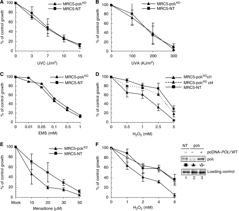
Figure 2.
Sensitivities of MRC5-polιKD cells to killing by various DNA-damaging agents. For each survival experiment, cells were exposed to increasing doses of the indicated genotoxic agent and counted 72 h later. Percentage of control growth was plotted for each data point, representing the mean of three independent experiments. (A) Cells were exposed to UVC, (B) UVA and (C) EMS for 1 h at 37°C in serum-free medium. (D) Cells were exposed to H2O2 for 10 min at 4°C. (E) Cells were exposed to menadione for 1 h at 37°C. (F) On the left panel, MRC5-polιKD cells were transfected with either an empty vector (closed triangle) or a vector expressing polι (open triangle). After 24 h, cells were exposed to H2O2 as described above. On the right panel, western blots of extracts of MRC5-polιKD and MRC5-NT cells containing an empty vector or polι expression vector, which were separated by SDS–PAGE and blotted using anti-polι antibody, are shown.

