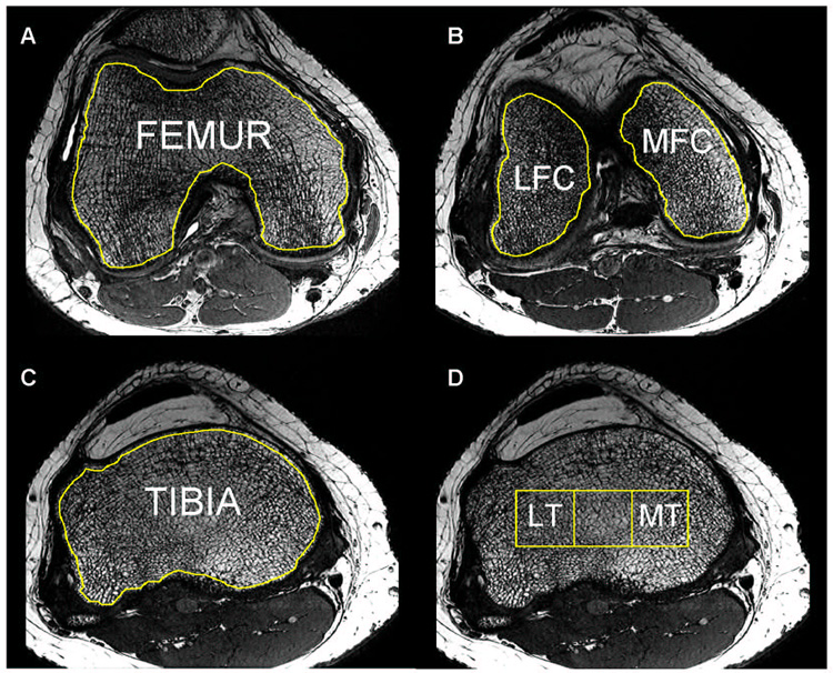
Figure 1.
Trabecular bone structure post-processing: bone and marrow regions of interest (ROI) were outlined for the femur (A), lateral and medial condyles (B), tibia (C) and lateral and medial tibia (D); the tibial grid was derived from the epicondylar distance (Unit [mm] = Epicondylar Distance × 100/9).

