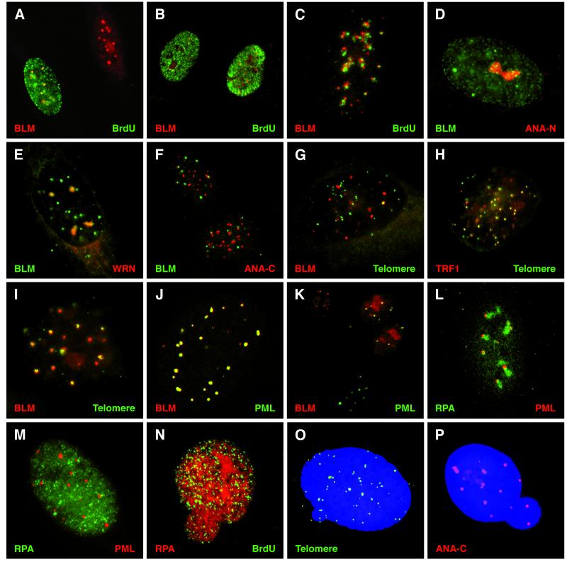
Figure 1.
Localization of α-BLM and sites of DNA replication, repeated sequence elements, and ND10s. (A–C) Normal human diploid fibroblasts (HG2619 or WI38) double-labeled with α-BLM (α-rabbit TR/2°) and α-BrdUrd mouse (α-mouse FITC/2°). Representative BrdUrd patterns are shown: pattern 3, middle to late S phase (A); patterns 1 and 2, early S phase (B); pattern 4, late S phase (C); pattern 5, small numbers of foci (not shown). (D) HG2619 cell stained with α-BLM (α-rabbit FITC/2°) and α-nucleolar human autoimmune sera ANA-N (α-human TR/2°). (E) HG2619 cell stained with α-BLM (α-rabbit FITC/2°) and α-WRN (α-rabbit TR/2°). (F) HG2619 cells stained with α-BLM (α-rabbit FITC/2°) and α-centromere human autoimmune sera ANA-C (α-human TR/2°). (G) WI38 cell stained with α-BLM (α-rabbit TR/2°) and hybridization of a DIG-labeled telomeric sequence probe (α-DIG FITC/2°). (H) SV40-transformed-BS fibroblast cell (HG2522) stained with α-TRF1 rabbit (TR α rabbit/2°) and hybridization of a DIG-labeled telomeric sequence probe (α-DIG FITC/2°). (I) SV40-transformed normal fibroblast cell (HG2855) stained with α-BLM (α-rabbit TR/2°) and hybridization of a DIG-labeled telomeric sequence probe (α-DIG FITC/2°). (J) Confocal image of a normal cell (HG2619) stained with α-BLM (α-rabbit TR/2°) and α-PML (α-mouse FITC/2°). (K) WI38 cells stained with α-BLM (α-rabbit TR/2°), α-PML (α-mouse FITC/2°). (L) Confocal image of HG2619 cell stained with α-RPA (α-rabbit FITC/2°) and α-PML (α-mouse TR/2°). (M) BS cell (HG2940) stained with α-RPA (α-rabbit FITC/2°) and α-PML (α-mouse TR/2°). (N) BS cell (HG3002) stained with α-RPA (α-rabbit TR/2°) and α-BrdUrd (α-mouse FITC/2°). (O) BS cell (HG3005) stained with 4′,6-diamidino-2-phenylindole and hybridization of a DIG-labeled telomeric sequence probe (α-DIG FITC/2°). (P) BS cell (HG2940) stained with 4′,6-diamidino-2-phenylindole and α-ANA-C (α-human TR/2°).

