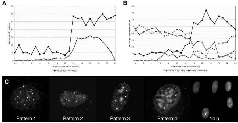
Figure 2.
Patterns of α-BLM staining during the cell cycle. Normal human diploid fibroblasts (HG2619) were serum-starved for 48 h and returned to growth by serum addition. Cells were harvested every hour and stained with α-BLM (α-rabbit TR/2°). Slides were pulse-labeled with BrdUrd and stained with α-BrdUrd (α-mouse FITC/2°). (A) Cells on slides were observed and counted for α-BLM staining and BrdUrd incorporation. (B) Distribution of α-BLM staining patterns as a function of time after serum addition. (C) Representative BLM patterns and a field of cells at 14 h after serum addition stained with α-BLM (α-rabbit TR/2°).

