Figure 4.

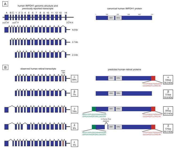
Human IMPDH1 transcripts and proteins. (A) Genomic structure and three IMPDH1 transcripts originally reported by Gu et al.17 Data suggested that all three transcripts encoded the canonical IMPDH1 protein shown on the right. (B) Observed retinal transcripts and predicted proteins. Transcripts α and β are from the exon 1F to 3′UTR R product subcloning. Transcripts γ, δ, and ε are from the exon A F to 3′UTR R product subcloning. Transcript percentages reflect results of separate subcloning experiments. Only transcripts seen in greater than 2% of clones from each subcloning experiment are reported. Green: novel protein regions from exon A. Red: residues resulting from exon 13b.
