
Figure 2.
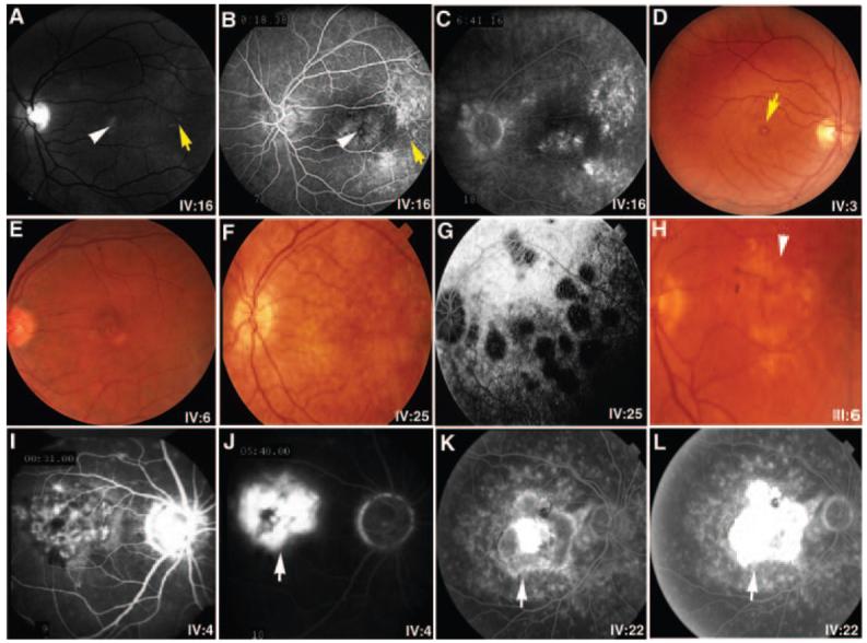
An assortment of representative fundus photographs and fluorescein angiograms from patients with maculopathy in SUNY901. Monochromatic fundus photographs (A) as well as the early (B) and late (C) fluorescein angiographic frames highlight the subtle features of early atrophic disease, including pigmentary changes (white arrowheads) and drusen (yellow arrows) in the macula of IV:16. (D, E) Fine yellowish drusenlike substance at the level of the retinal pigment epithelium with a small yolklike lesion (yellow arrow) in the foveas of IV:3 and IV:6, respectively. Photograph (F) and the late angiographic frame (G) show pigment epithelial atrophy and scalloped areas of choriocapillaris loss in later stages of atrophy in IV:25. (H) Extensive geographic atrophy of central macula encountered in III:6 is shown. Early (I, K) and late (J, L) angiographic frames highlight features of the exudative disease, showing subfoveal hyperfluorescence and leakage attributable to an ill-defined choroidal neovascular membrane in IV:4 (I, J) and a disciform scar in IV:22 (K, L). The choroid is especially dark in IV:4. White arrows: Choroidal neovascularization is indicated.