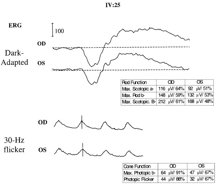
Figure 3.
Full-field ERG in patient IV:25. Dark-adapted (scotopic) and light-adapted (photopic) ERGs were recorded with bipolar Burian Allen contact lens electrodes in accordance with the 1999 International Society for Clinical Electrophysiology of Vision (ISCEV) standards. Representative responses under maximum (A) scotopic and (B) photopic conditions are shown. The included tables list absolute peak wave amplitudes for the maximum scotopic stimulus (threshold +4 log); scotopic a wave threshold (max. rod b) and maximum photopic responses. Percentages represent the fraction of the lower limit of normal (mean, −2.5 sec/d) for the listed stimulus condition.

