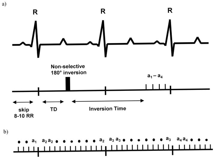
Figure 1.

(a.) Timing of the IR-sequence, where 8–10 R-R intervals are skipped between each acquisition. Due to the animal's high heart rate, the readout occurs 1–2 heart beats after the inversion pulse. The imaging portion of the sequence consists of 4 phase encoding steps (a1–a4) timed to end diastole. (b.) The T1-CF sequence can be repeated for each heartbeat. Only one phase encoding step per cardiac phase is acquired during each R-R interval.
