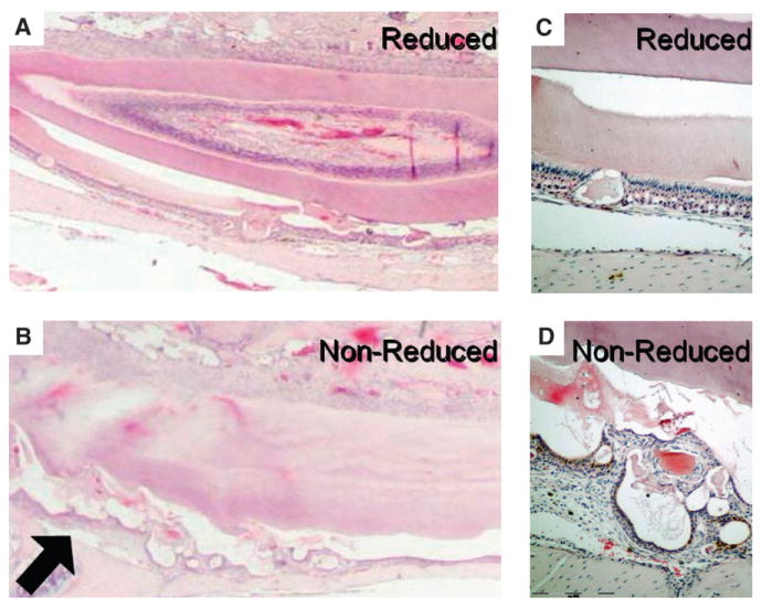
Figure 7.

Partial rescue of the ameloblast layer by removal of occlusal force. A) Trimmed incisor from the knock-out mouse. Note the fairly homogeneous enamel surface as well as the ameloblast layer. B) The untrimmed side showing increased cyst formation (arrow). C) Higher magnification of the ameloblast layer on the trimmed side showing one of the small defects. D) Higher magnification of the irregular and detached ameloblast layer on the untrimmed side. (H&E staining: A through D; original magnification: A and B, ×1; C and D, ×10).
