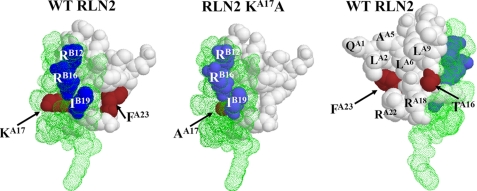
FIGURE 8.
Comparative structure modeling of mutant peptides based on the crystal structure of human RLN2. Three-dimensional structure of the wild type human RLN2 and the KA17A mutant based on the RLN2 crystal structure (PDB 6RLN). The wild type RLN2 structure is shown on the left and right panels. The KA17A mutant is shown on the middle panel. The A and B chains are indicted by white filled space and green dot space, respectively. The amino acids at A16, A17, and A23 positions are indicated by the brown filled space. The ArgB12, ArgB16, and IleB19 residues in the RXXXRXXI motif of B chain are represented by the blue filled space.

