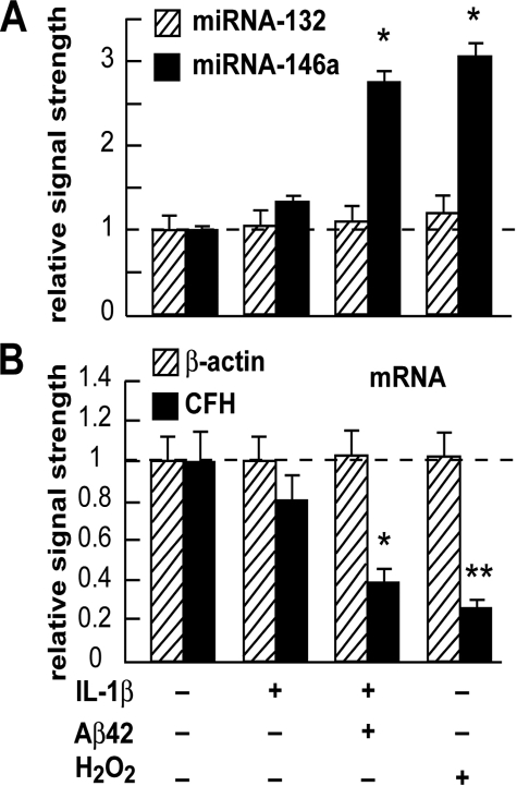
FIGURE 2.
Specific up-regulation of miRNA-146a, but not miRNA-132 in IL-1β, IL-1β+Aβ42, and in H2O2 treated HN cells in primary culture. A, 2-week-old HN cells were treated with IL-1β (10 ng/ml cell medium), IL-1β+Aβ42 (8 μm) (22), and/or H2O2 (2.5 μm) in HN maintenance medium for 1 week, representing, respectively, inflammatory cytokine IL-1β-, Aβ42 peptide-, and/or ROS-mediated stress for one-third of their in vitro lifetime. Total small RNA was isolated, radiolabeled, and used to probe dot blot arrays containing brain-enriched miRNA targets as previously described (20, 26). The results indicate a specific up-regulation of miRNA-146a but not a related brain-enriched miRNA-132, after IL-1β, IL-1β+Aβ42, or H2O2 treatment, to 1.4-, 2.6-, and 3.1-fold, respectively, over untreated normally aging HN cell controls. Control miRNA levels (miR-132 and miR-146a) arbitrarily set to 1.0 (dashed horizontal line; n = 4). *, p < 0.05 (ANOVA). B, down-regulation of CFH mRNA in the same HN cell samples as in A. Taken together with the data shown (A), the results suggest that miRNA-146a is up-regulated in HN cells stressed with inflammatory cytokines and amyloid peptides or oxidative stressors known to be present in AD brain (2–6, 14, 26).

