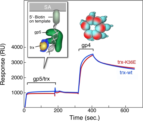
FIGURE 6.
Binding of gp4 to gp5-trx complexes bound to DNA. A primer-template with biotin at the 5′-end of the template strand is immobilized on an SA-sensor chip. gp5/trx-wt or gp5/trx-K36E is injected, followed by gp4. Binding studies were carried out as described under “Experimental Procedures.” One hundred response units of the biotinylated primer-template is coupled to the surface. gp5/trx-wt or gp5/trx-K36E is injected at concentrations of 0.2 μm gp5 and 8 μm trx and trx-K36E (ratio 1:40) in flow buffer containing 1 mm dTTP and 10 μm ddGTP, at saturating conditions of gp5/trx and primer-template. The 100 RU resulting from the coupling of primer-template is subtracted from base line. gp4 is injected at a concentration of 0.7 μm (monomer) in flow buffer containing 0.1 mm ddGTP and 2 mm dTTP. The start and end of the injections are indicated. Red indicate complexes, with trx-K36E and blue indicate complexes with trx-wt.

