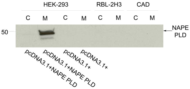
Figure 6. NAPE PLD is not detectable in RBL-2H3 cells or CAD cells.
HEK-293 cells, transfected either with pcDNA3.1+ NAPE PLD or vector pcDNA3.1+, and RBL-2H3 cells were lysed on ice for 10 min in lysis buffer. The whole cell lysates were then subjected to separation into the cytosolic and membrane fractions as described in Materials and Methods. The samples were then resolved on SDS PAGE and transferred onto a polyvinylidene difluoride membrane. The presence of NAPE PLD (46 kDa) was revealed using the rabbit anti-NAPE PLD polyclonal primary antibody (1:500, Cayman Lipids) and a goat anti-rabbit (1:2000), horseradish peroxidase-labeled secondary antibody followed by an exposure with ECL detection reagents. Data are representative of three separate experiments.

