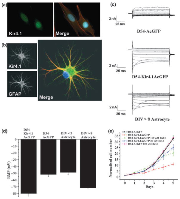
Fig. 5.
Role of Kir4.1 in glial growth control: (a) Immunostaining with antibodies to Kir4.1 and the cytoskeleton with Phalloiden (Kir4.1: green, Phalloiden:red) show Kir4.1 labels the cell nucleous. (b) In contrast in astrocytes Kir4.1 (green) labels the cell membrane (the cytoskeleton is labeled with GFAP, red). (c) This difference is also reflected in patch-clamp recordings from another glioma cells line (D54) which lack inward currents compared to SC astrocytes that express prominent inward currents. However, upon transfection of D54MG cells with a GFP containing plasmid encoding Kir4.1, these glioma cells express inward currents that are indistinguishable from astrocytic Kir currents. (d) Expression of Kir4.1 in glioma caused their resting potential to shift ~30 mV negative making it similar to that of differentiated astrocytes. (e) The presence or absence of functional Kir4.1 channels determined the rate of growth of D54 glioma cells, in which growth was retarded when Kir4.1 channels were functional. However, depolarizing the cell with 20 mM K+ was sufficient to rescue growth even when Kir4.1 was over-expressed [(a) With permission from Olsen and Sontheimer 2004a; (b) With permission Olsen et al. 2006; (c–e) With permission from Higashimori and Sontheimer 2007].

