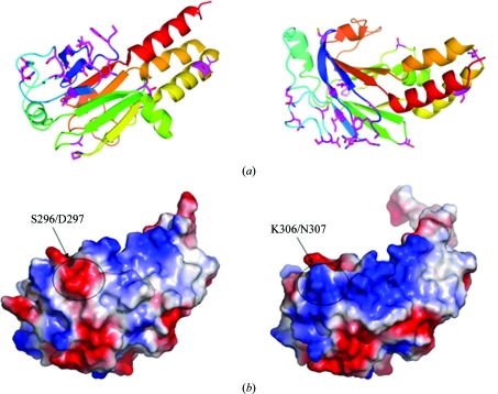
Figure 3.
Structural features of Mad MH2. (a) The variant residues of Mad MH2 indicated in Fig. 2 ▶ are coloured magenta. The two views of the structure are related by a 90° rotation around a horizontal axis. (b) Surface representations of Mad MH2 (left) and Smad1 MH2 (right), coloured according to electrostatic potential, with positive and negative charges in blue and red, respectively.

