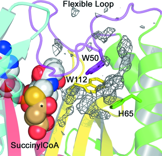
Figure 5.
Secondary binding groove. Residual electron density (final F o − F c contoured at 2.5σ) in a secondary binding groove adjacent to the SucCoA-binding site. Conserved binding residues are shown as sticks. SucCoA is shown as CPK spheres, with the succinyl moiety shown in brown C atoms and the acyl carbon marked with an asterisk.

