
FIG. 1.
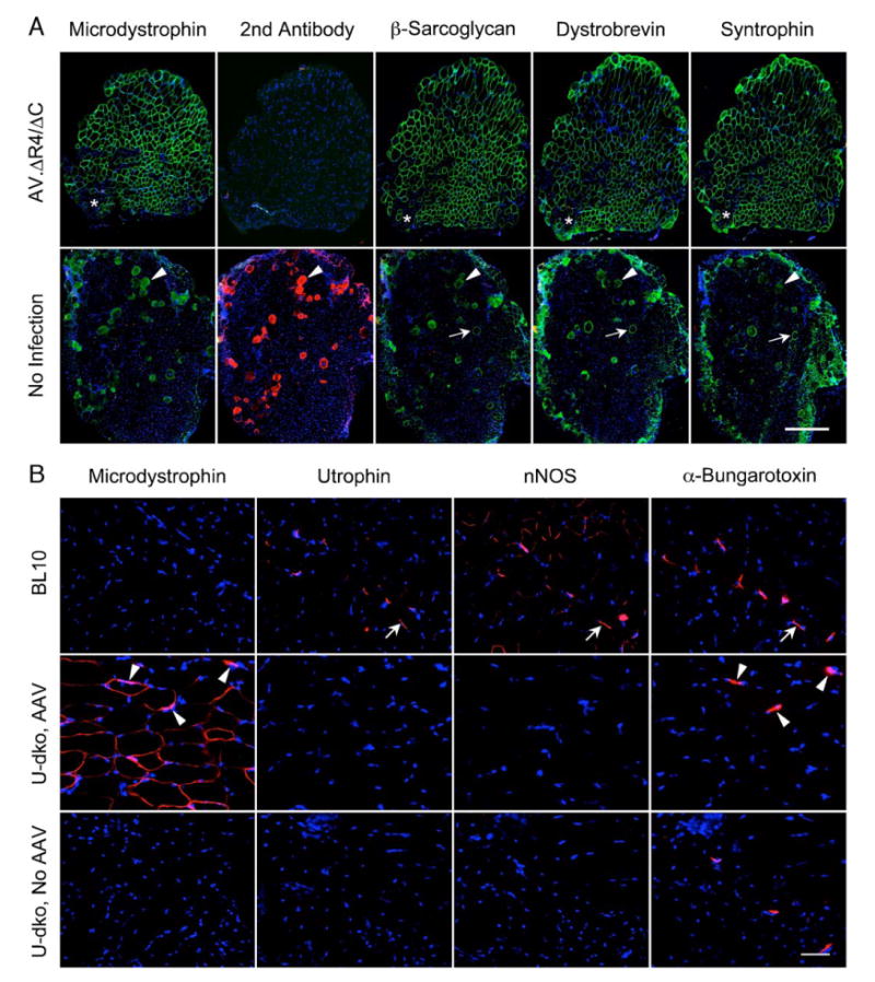
C-terminal-truncated microdystrophin restores the DGC, but not nNOS, in the sarcolemma of u-dko mice. 6 × 10¹⁰ vg particles of AAV-6 AV.ΔR4/ΔC were delivered to the anterior compartment of the left hind limb in 3-day-old neonatal u-dko mice. The contralateral limb was injected with an equal volume of saline buffer. The DGC components were examined by immunostaining at 9 weeks after gene transfer. (A) Representative photomicrographs of u-dko muscle serial sections immunostained with monoclonal antibodies for the dystrophin N-terminal domain, β-sarcoglycan, dystrobrevin, and syntrophin, respectively. Secondary antibody alone (exactly the same antibody as used for other immunostainings except it is conjugated to Alexa 594) is included as a negative control for nonspecific staining. Secondary antibody alone also reveals immunoglobulin uptake in injured myofibers. Nuclei are revealed by DAPI staining. Asterisk, a region not transduced by AAV; arrow, a revertant myofiber; arrowhead, an injured myofiber with sarcolemma leakage. Scale bar, 300 μm. (B) Representative photomicrographs of serial sections immunostained with monoclonal antibodies for the dystrophin N-terminal domain (specific for AV.ΔR4/ΔC) and utrophin and with polyclonal antibody for nNOS. Neuromuscular junctions are revealed with Alexa 594-conjugated α-bungarotoxin. Nuclei are revealed by DAPI staining. Arrow, expression of utrophin and nNOS at the neuromuscular junctions in BL10 muscle; arrowhead, microdystrophin expression is enhanced at the neuromuscular junctions in AV.ΔR4/ΔC-infected u-dko muscle. Scale bar, 50 μm.