
Figure 3.
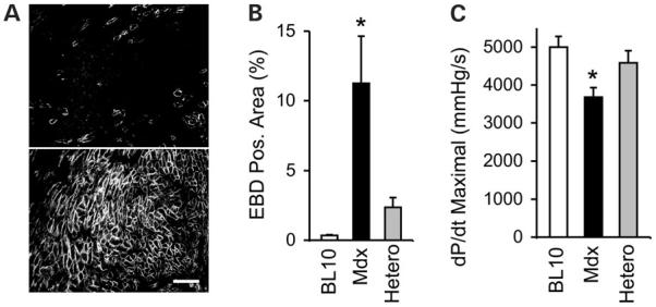
Mosaic dystrophin expression in heterozygous mdx mice protects the heart from stress-induced cardiomyopathy. (A) Dystrophin expression in the heart of heterozygous female mice. Top and bottom panels depict regions of low and high dystrophin expression, respectively. Scale bar: 100 μm. (B) Quantification of sarcolemma damage (EBD positive area) in the hearts of different mouse strains following β-isoproterenol challenge. (C) Correction of hemodynamic defect in heterozygous mice. Bar graph shows the dP/dt maximal. Asterisk denotes results in mdx were significantly different from these in BL10 or heterozygous mice. EBD, Evans blue dye; Hetero, heterozygous mice. Adapted from Yue et al. (2004) Hum. Mol. Genet., 13, 1669.