
Figure 1. Wide-spread transduction of skeletal muscle following intravenous delivery of trans-splicing AAV vectors in newborn mice.
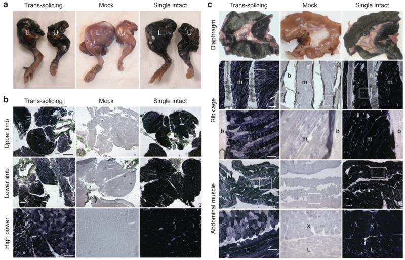
Representative histochemical staining photomicrographs from each group (N = 3–5 mice/group) are shown. (a) Full-view of the whole mount limb muscle (L, lower limb; U, upper limb). (b) Cross-sections of the entire limb (top two rows; bar, 1 mm) or high magnification view of representative areas of muscles in the lower limb (bottom row; bar, 200 μm), respectively. Dotted lines in low magnification photomicrographs mark the location of bone. (c) AP expression in respiratory muscles including the diaphragm (top row, whole mount images), inter-costal muscles (middle two rows, cross sections), and abdominal muscles (bottom two rows, cross-sections), respectively. Photomicrographs in high magnification (bar, 100 μm) are the closer view of the boxed areas in the respective low magnification (bar, 400 μm) photomicrographs. b, bone (rib); m, muscle; X, regions where myofibers are in cross-sectional orientation; L, regions where myofibers are in longitudinal orientation.