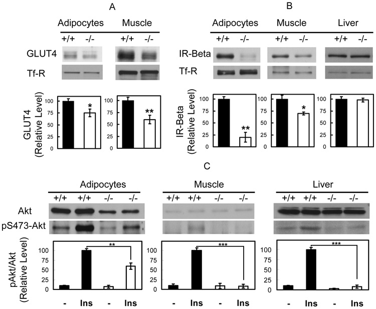
Figure 4.
GLUT4 expression is reduced and Insulin signaling is impaired in Cavin null mice. After insulin injection (i.p. 0.75 U/kg), animals were sacrificed and whole cell lysates of wild type (Cavin+/+) and Cavin knockout mice (Cavin−/−) (16 weeks old strain-matched males) from adipocyte, muscle and liver tissues were prepared in RIPA buffer. Representative western blots and quantitative bar graphs are shown for the detection of (A) GLUT4, (B) IR-beta protein levels and (C) total Akt and Akt S473 phosphorylation levels. The statistical values are displayed as means and SE of 3 independent experiments. (*P < 0.05, **P < 0.01, ***P < 0.001).

