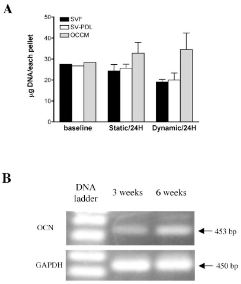
Figure 1.

(A) Cell attachment efficiency to PLGA scaffolds under static and dynamic culture conditions. After 24 h culture (compared to baseline DNA content) in static conditions, SV-F, SV-PDL, and OCCM cells attached to PLGA scaffolds by 88.5%, 95.9%, and 115.5%, respectively, whereas after dynamic culture for 24 h, SV-F, SV-PDL, and OCCM cells attached to PLGA scaffolds by 74.9%, 69.0%, and 121.5%, respectively. (B) Cementoblasts maintain osteocalcin gene expression in 3D PLGA scaffolds. Photograph of 2% agarose gel electrophoresis of RT-PCR products of mRNA extracted from OCCM cells cultured in dynamic conditions in spinner flasks for 3 and 6 weeks shows that osteocalcin expression was detected at both 3 and 6 weeks. GAPDH was used as a control.
