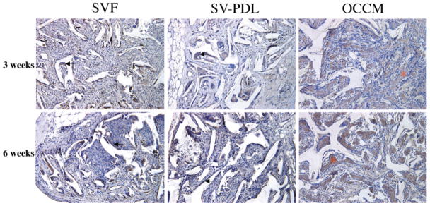
Figure 4.
Immunodetection of BSP in cell-PLGA constructs. Rabbit antimouse sialoprotein antibody was used to detect BSP irnmunoreaction in 3-and 6-week specimens. In samples from both 3 and 6 weeks, SV-PDL/PLGA and SV-F/PLGA scaffold positive staining for BSP was noted in multinuclear cells and spindle-shaped cells (solid arrows). Strong BSP staining was noted in the mineralized extracellular matrix of samples obtained from OCCM/PLGA implants (*). This is a representative experiment, with similar results noted on two occasions. Antibody control specimens (no primary antibody) were devoid of staining (data not shown). (Hematoxylin counterstain, 20× original magnification)

