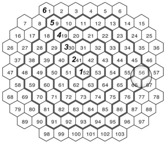
Fig. 1.

Hexagonal areas and borders of ring average patterns. The numbers inside the hexagonal areas indicate the stimulated areas provided by the VERIS system (field view). The outside borders of the ring average patterns commonly used in clinical practice are denoted by bold contours and designated by bold numerals. The gray circle indicates the approximate location of the optic nerve head
