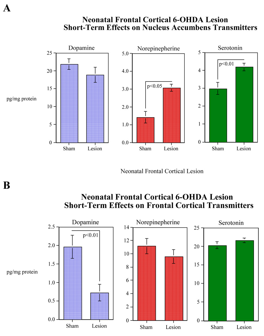
Figure 1.
Effects of neonatal 6-OHDA lesion of the frontal cortex on levels of dopamine, norepinepherine and serotonin in the frontal cortex (Panel A), and dopamine, norepinepherine and serotonin in the nucleus accumbens (Panel B) 10 days following lesioning (mean±sem). The figure depicts pooled data from both male and female rats. Control animals were infused with artificial an equal volume of artificial cerebral spinal fluid. N=8 for all neurotransmitters in both treatment conditions except for the cortical norepinephrine in the sham group which is 7.

