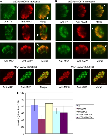
Figure 4.
Functional characterization of the mic6 knock out strain and mic6 knock out parasites complemented with either full-length TgMIC6 or ΔEGF2-MIC6TY and ΔEGF3-MIC6TY mutant proteins. Immunofluorescence assays showing that (A) ΔEGF2-MIC6TY and (B) ΔEGF3-MIC6TY are stably expressed and correctly targeted to the micronemes (anti-TY; green). Transport of TgMIC1 to the micronemes is partly restored in these parasites (anti-MIC1; green), as shown by colocalization with the microneme marker AMA1 (anti-AMA1; red). White arrowheads indicate examples of correct targeting to the micronemes. Note that ΔEGF2-MIC6TY is TY-tagged. A control assay is shown (bottom in A,B) in which MIC1-ΔGLD is unable to interact with either EGF domain from MIC6 and rescue correct microneme localization. TgMIC6 and TgMIC1 are retained in the endoplasmic reticulum/Golgi and dense granules, respectively. A green/magenta version of this figure is shown as supplementary Fig 4 online. (C) Functional assay comparing various parasite strains for their invasion efficiency using RH-2YFP parasites as an internal standard for parasite fitness and human foreskin fibroblasts as host cells. Invasion data were compared by one-way ANOVA followed by a Newman–Keuls test. Invasion data for ΔEGF2-MIC6/6 and ΔEGF3-MIC6/6 parasites are statistically different from MIC6KO and RH-2YFP parasites, supporting the partial complementation of invasion activity in these mutants. ANOVA, analysis of variance; EGF, epidermal growth factor-like; GLD, galectin-like domain; ko, knock out; MIC, microneme protein; RH-2YFP, RH strain of Toxoplasma gondii transformed with a plasmid expressing yellow fluorescent protein.

